Historically serum creatinine was the lab value used to assess kidney function. Answers after the references.

Illustration Of Dw Mri Signal Decay Of Various Types Of Tissues With Download Scientific Diagram
GadoliniumIII containing MRI contrast agents often termed simply gado or gad are the most commonly used for enhancement of vessels in MR angiography or for brain tumor enhancement associated with the degradation of the bloodbrain barrier.
. The physics of MRI are complicated and much harder to understand than those underpinning image generation in plain radiography CT or ultrasound. This section of the website will explain planning for various types of MRI scans MRI protocols positioning for MRI and common indications for MRI scans. Basically at 3 T it will be twice the value obtained at 15 T.
It is important to read this manual before conducting an MRI scan on a patient with an. For large vessels such as the aorta and its. Although sonography is the primary imaging test for a palpable thyroid nodule or known thyroid malignancy thyroid abnormalities are frequently first discovered on other cross-sectional modalities of computed tomography CT and magnetic resonance imaging MRIA thyroid lesion may be seen as an incidental finding or CT and MRI.
As a result the T1 value of bound or structured water is much less than free water 400800 msec4 6 Fat typically has a short T1 value. An arthrogram is a series of images of a joint after injection of a contrast medium usually done by fluoroscopy or MRIThe injection is normally done under a local anesthetic such as Novocain or lidocaineThe radiologist or radiographer performs the study using fluoroscopy or x-ray to guide the placement of the needle into the joint and then injects around 10 ml of contrast based on age. EGFR takes into account the serum creatinine value and also patient age race and gender which affect kidney function results.
When the calculator has enough information the O-RADS MRI Risk Score will display. Do not scan patients in a 3 T magnetic field with a B1RMS value 28 µT when the isocenter center of the MRI bore is inferior to the C7 vertebra. Several pathologic conditions may manifest as an osteochondral lesion of the knee that consists of a localized abnormality involving subchondral marrow subchondral bone and articular cartilage.
MAGNETIC RESONANCE IMAGING WITHOUT CONTRAST FOLLOWED BY WITH CONTRAST BREAST. AZURE MRI SURESCAN ASTRA MRI SURESCAN. Additional preparation sequences can also be performed to manipulate the magnetization and so the image contrast eg.
Multiple choice questions True TFalse F. Presence of peritoneal mesenteric or omental nodularity or. At UCSF we use this very accurate.
A scan above 28 µT may increase the. Even when I was an office of one I never ever felt alone. Toxicol Ind Health 20112730713.
A the investment MRI pours into its offices and b the camaraderie between owners. A better and more accurate measure is a lab result called estimated glomerular filtration rate eGFR. B and C In accordance with the Standardization of Acquisition and Post-Processing study combined protocol of dynamic contrast-enhanced DCE MR perfusion transfer constant map B was obtained first with 005 mmolkg gadobutrol at 2 mLs and 20 mL saline flush followed by dynamic susceptibility contrast-enhanced DSC MR perfusion imaging.
Fee schedules relative value units conversion factors andor related components are not assigned by the AMA are not part of CPT and the AMA is not recommending their use. Although understanding of these conditions has evolved substantially with the use of high-spatial-resolution MRI and histologic correlation it is impeded by inconsistent. Do not scan patients in a 3 T magnetic field with a B1RMS value 28 µT when the isocenter center of the MRI bore is inferior.
This page will explain more about MRI brain. Press the reset button in the upper right corner after each lesion to reset the calculator. Over 450 million doses have been administrated worldwide from 1988 to 2017.
I joined MRI because they offer tremendous training tools and a group of recruiting firms that often feels more like a family than a network. Magnetic resonance imaging and safety aspects. ICD-10-CM Codes that Support Medical.
I get the most value from. Academic Radiology publishes original reports of clinical and laboratory investigations in diagnostic imaging the diagnostic use of radioactive isotopes computed tomography positron emission tomography magnetic resonance imaging ultrasound digital subtraction angiography image-guided interventions and related techniques. BILATERAL CPTHCPCS Modifiers NA.
On the MRI SureScan mode allows the patient to be safely scanned while the device continues to provide appropriate pacing.
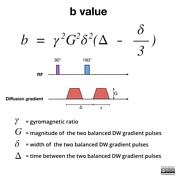
2 Examples Of Diffusion Weighted Images For Different B Values The Download Scientific Diagram

Pdf Diffusion Weighted Mri And Optimal B Value For Characterization Of Liver Lesions
How Does B Value Affect Hardi Reconstruction Using Clinical Diffusion Mri Data Plos One

Diffusion Mri Signal Decay Versus B Value A The Diffusion Signal Download Scientific Diagram

Figure 3 From Zafer Koc Value For Characterization Of Liver Lesions B Diffusion Weighted Mri And Optimal Semantic Scholar

Advantage Of High B Value Diffusion Weighted Imaging For Differentiation Of Common Pediatric Brain Tumors In Posterior Fossa European Journal Of Radiology

Conventional Mri Dwi And Adc Images Of Braf V600e Mutant And Download Scientific Diagram
How Does B Value Affect Hardi Reconstruction Using Clinical Diffusion Mri Data Plos One
Fig 3 High B Value Diffusion Weighted Mr Imaging Of Adult Brain Image Contrast And Apparent Diffusion Coefficient Map Features American Journal Of Neuroradiology

Diffusion Tensor Imaging Dti Fiber Tracking Imagilys

High B Value Diffusion Weighted Mr Imaging Of Normal Brain At 3t European Journal Of Radiology

Illustration Of The Signal Decrease When The Echo Time And The B Value Download Scientific Diagram
High B Value Diffusion Weighted Mr Imaging Of Suspected Brain Infarction American Journal Of Neuroradiology

Diffusion Weighted Imaging Radiology Reference Article Radiopaedia Org

Diffusion Weighted Imaging Radiology Reference Article Radiopaedia Org
Fig 4 High B Value Diffusion Weighted Mr Imaging Of Suspected Brain Infarction American Journal Of Neuroradiology

Physical Principles Use Of High B Values And Clinical Applications Of Diffusion Weighted Imaging Of Ischemic Stroke

Signal Intensity Changes With Increasing B Values A 29 Year Old Female Download Scientific Diagram

Combining Perfusion And High B Value Diffusion Mri To Inform Prognosis And Predict Failure Patterns In Glioblastoma International Journal Of Radiation Oncology Biology Physics
